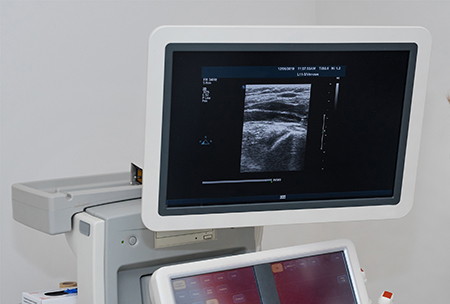
The color doppler ultrasound allows to identify the venous insufficiency to the legs. This technique is mainly used to diagnose if a leg edema is caused by a venous disease and the possible remedies that can be applied. It allows the origin and characteristics of the venous insufficiency to be identified individually and serves to guide the necessary treatment for each patient.
Doppler ultrasound is a non-invasive test that calculates the flow of blood in the blood vessels by bouncing high frequency sound waves off the circulating red blood cells. In standard ultrasound, sound waves are used to create images, but the blood flow cannot be shown. Doppler ultrasound can help diagnose many conditions, including the following:
blood clots
Poorly functioning valves in the leg veins, which can cause blood or other fluids to pool in the legs (venous insufficiency).
Heart valve defects and congenital heart disease.
A blocked artery (arterial occlusion).
Reduced blood flow to the legs (peripheral arterial disease).
Widened arteries (aneurysms).
Narrowing of an artery, such as in the neck (carotid artery stenosis).
Doppler ultrasound can calculate the speed of blood flow by measuring the proportion of changes in blood tone (frequency). During Doppler ultrasound, a specialized ultrasound imaging technician (sonographer) presses a small handheld device (transducer) about the size of a bar of soap onto the skin of the body part being examined and moves it as needed.
This test can be performed as an alternative to more invasive procedures, such as arteriography, which involves injecting a dye into the blood vessels to make them clear on the x-ray. It can also help the health care provider detect injury to the arteries or monitor certain treatments in the veins and arteries.